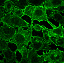
Microsome Preperation
- Cells are grown to confluency on P150 dishes.
- Place dishes on ice and proceed to do the rest of the protocol on ice. It is possible to process 5 plates simultaneously.
- Wash cells with ice cold PBS 2X by flooding the plate and decanting off wash
- Wash cells 1X with ice cold lysis buffer by flooding the plate and decanting off wash.
- Remove any remaining wash with a 1 ml pipette and add 1 ml of Lysis buffer containing 2X protease inhibitors to each plate.
- Scrape the cells off the plate and place in Dounce homogenizer. Incubate 5 minutes on ice.
- Rupture cells with 5-10 strokes of the homogenizer. Ruptured nuclei will release chromatin that will bind up the microsomes and reduce yield. It is better to under-homogenize than over. 70% lysis is a good target.
- Efficiency is checked by examination of lysate stained with Trypan blue using inverted microscope.
- Osmolarity is balanced by adding 5X sucrose buffer to homogenizer and mixing with 1 stroke
- Cell debris is pelleted at maximum speed using clinical centrifuge at 4°C (3000 xg)
- Supernatant containing microsomes is spun at 16000 xg in refrigerated microfuge at 4°C for 30 minutes. Large volumes can be spun at 26000 rpm in ultracentrifuge using SW28 rotor
- Microsome pellet is resuspended in appropriate buffer for experiment.
- Bradford protein assay used to determine protein concentration.
- Samples are stored at -80°C
Lysis Buffer:
10 mM HEPES pH 7.2
1 mM EDTA
2x Protease Inhibitor cocktail
5X Sucrose Buffer:
1.25 M Sucrose
10 mM HEPES pH 7.2
1000X Protease Inhibitor Cocktail:
Leupeptin (Sigma L2884): 1mg/ml
Aprotinin (Sigma A1153): 2mg/ml
E64 (Sigma E3132): 3.57mg/ml
Benzamidine (Sigma B6506): 156.6mg/ml
Pefablock (Roche 1429876): 2M

